
Figure 1. Glipizide inhibits angiogenesis in embryonic CAM and YSM assays.
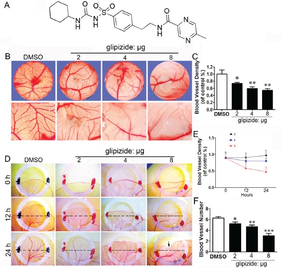
Chick CAM and YSM assays were used to determine whether glipizide can inhibit angiogenesis to suppress tumor growth. (A) Chemical structure of glipizide. (B) The upper panels show that the blood vessel plexus of 11-day-old chick embryos following treatment with 2, 4 and 8 μg of glipizide or DMSO. The lower panels show the appearance of blood vessel plexus at higher magnification as indicated by white dotted squares in upper panels. (C) The bar chart shows the relative blood vessel density on CAM following glipizide and DMSO treatment. (D) The entire egg content was transferred into a sterile petri dish after two-day incubation. The upper panels show the appearance of the blood vessel plexus at the start of experiment (0 h) for control and 2, 4 and 8 μg of glipizide. The middle and lower panels show the appearance of blood vessel plexus after 12 h and 24 h incubation, respectively. (E) Statistical chart shows the blood vessel density for control and glipizide treatment group. (F) Bar chart compares the number of blood vessels on YSM between control and glipizide treatment group. * p < 0.05; ** p < 0.01.