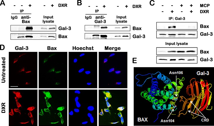
Fig.3. Gal-3 binds to Bax through CRD in response to apoptotic stimulus.
A and B, Co-immunoprecipitation assay. TPC1 cells were treated with 0.5 μM DXR for 24 hours. Cell lysates were immunoprecipitaed with IgG rabbit, polyclonal anti-Bax, or polyclonal anti-Gal-3 antibody. The immunoprecipitates and input lysates were analyzed by immunoblotting with indicated antibodies. Input lysates indicate lysates used for immunoprecipitation from TPC1 cells and were used as positive control. C, TPC1 cells were pretreated with 1% of GCS-100/MCP for 3 hours, and then either left untreated or treated with 1 μM DXR for 24 hours. Cell lysates were immunoprecipitated with polyclonal anti-Gal-3 antibody. The immunoprecipitates and input lysates were analyzed by immunoblotting with indicated antibodies. D, Co-localization of Gal-3 and Bax in TPC1 cells treated with 0.5 μM DXR for 24 hours. TPC1 cells were immunofluorescently labelled with anti-Gal-3 (red), anti-Bax (green) antibodies and Hoechst 33258 (nuclear stain, blue). Scale bar represents 50 μm. E, Prediction of the interaction of Gal-3 carbohydrate recognition domain (CRD) with Bax. The references about the structure of Gal-3 CRD and Bax were indicated in Materials and Methods. In silico docking was performed using Second ClusPro 2.0 server (http://cluspro.bu.edu/login.php). Asn means asparagine.

