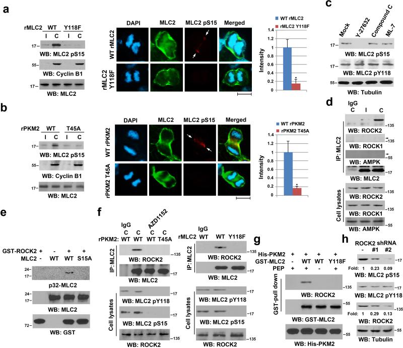
Figure 4. MLC2 pY118 Primes ROCK2-mediated MLC2 pS15.
Immunoblotting analyses were performed with the indicated antibodies. C, cytokinesis; I, interphase (no thymidine release).
(a, b) U87 cells with reconstituted expression of WT rMLC2, or rMLC2 Y118F (a), WT rPKM2, or rPKM2 T45A (b) were synchronized by double thymidine block (2 mM) for 43 h, with or without release for 10 h (left panel). Immunofluorescence analyses of cells in cytokinesis were performed with the indicated antibodies (middle panel). Scale bars, 10 μm. The relative fluorescence intensity of the indicated proteins in the contractile ring of 100 cells from each cell line was quantified. Data represent the mean ± SD of three independent experiments (right panel). *P<0.01: statistically significant value in relation to U87 cells with PKM2 depletion and reconstituted expression of WT rPKM2, Student's t-test.
(c) U87 cells, synchronized by double thymidine block (2 mM) for 43 h, were released for 9 h, followed by MG132 (25 μM) treatment for 1.5 h to arrest cells at metaphase. MG132 was then replaced with Y-27632 (10 μM), compound C (20 μM), or ML-7 (10 μM) for 30 min before cell harvesting.
(d) U87 cells were synchronized by double thymidine block (2 mM) for 43 h, followed by no release or release for 10 h. Immunoprecipitation analyses were performed.
(e) In vitro kinase assays were performed by mixing purified recombinant active GST-ROCK2 with purified PKM2-phosphorylated MLC2 for autoradiography.
(f) U87 cells with depleted PKM2 or MLC2 and reconstituted expression of WT rPKM2 or rPKM2 T45A (left panel), WT rMLC2 or rMLC2 Y118F (right panel) were synchronized and arrested at metaphase, as described in (c). MG132 was then replaced with AZD1152 (100 nM) for 30 min before cell harvesting (left panel). Immunoprecipitation analyses were performed.
(g) WT GST-MLC2 on agarose beads was incubated with purified Aurora B-phosphorylated His-PKM2 protein for a kinase assay in the presence or absence of PEP, which was followed by incubation with the lysate of HeLa cells synchronized by double thymidine block (2 mM) for 43 h and release for 10 h.
(h) U87 cells that expressed a control or two different ROCK2 shRNAs were synchronized by double thymidine block (2 mM) for 43 h and release for 10 h.

