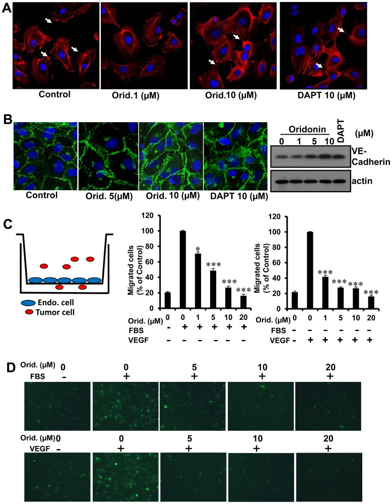
Figure 5. Oridonin increased the cell-cell connections of HUVECs and decreased tumor cell transendothelial invason.
(A) HUVEC morphology and cell-cell contacts by immunofluorescence assay. Arrows show the contact of endothelial cell edge with different concentrations of Oridonin. (B) HUVECs were treated with Oridonin or DAPT for 24 hours. Cells were fixed and stained with VE-cadherin. Photographs were obtained through a confocal microscope (left panel). HUVECs were treated with Oridonin or DAPT for 24 hours, and cells were harvested. VE-cadherin expression was examined by western blot analysis (right panel). (C) Transendothelial migration of 4T1 breast tumor cells with FBS or VEGF stimulation of HUVECs. 2×10⁵ HUVECs were grown to confluency for 48 hours on the Transwell membrane. HUVECs were treated with FBS or VEGF (20 ng/ml) for 18 hours. 1×10⁵ 4T1-GFP cells serum-starved overnight were added on the monolayer of HUVECs and incubated for 6 hours. After migrated cells were fixed and stained, the photographs were acquired. (D) The represent photographs of Fig. 5C. (*, P<0.05; **, P<0.01; ***, P<0.001).

