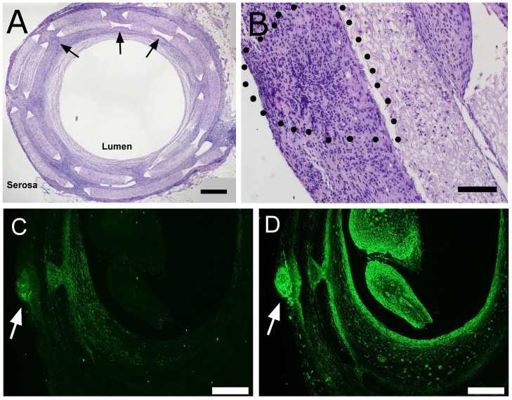
Figure 12. Histological analysis of implant sections.
Scaffolds with either GFP-expressing ISMC or MS were retrieved after two weeks in vivo and were visualized in cross-section with H&E staining (A, B). A representative image of an ISMC-seeded explant (A, 40x, 500-µm scale) shows laser-cut pores (arrows) to improve cellular and vascular infiltration through dense PCL scaffold (PCL) from the “serosal” to luminal side of the implant. GFP-expressing MS (B, 200x, 100-µm scale) on the “serosal” surface of a PCL implant is outlined with a dotted line. Scaffolds with GFP-expressing MS were immunofluorescently labeled with antibodies to GFP or SMA (C, D). GFP-expressing and MS (C) survived the 2 week implantation and migrated out from the MS. Non-GFP-expressing cells from the host also permeated the PCL implant and had SMA immunofluorescence (D). 40x magnification, 500-µm scale bar.

