Figure 1.
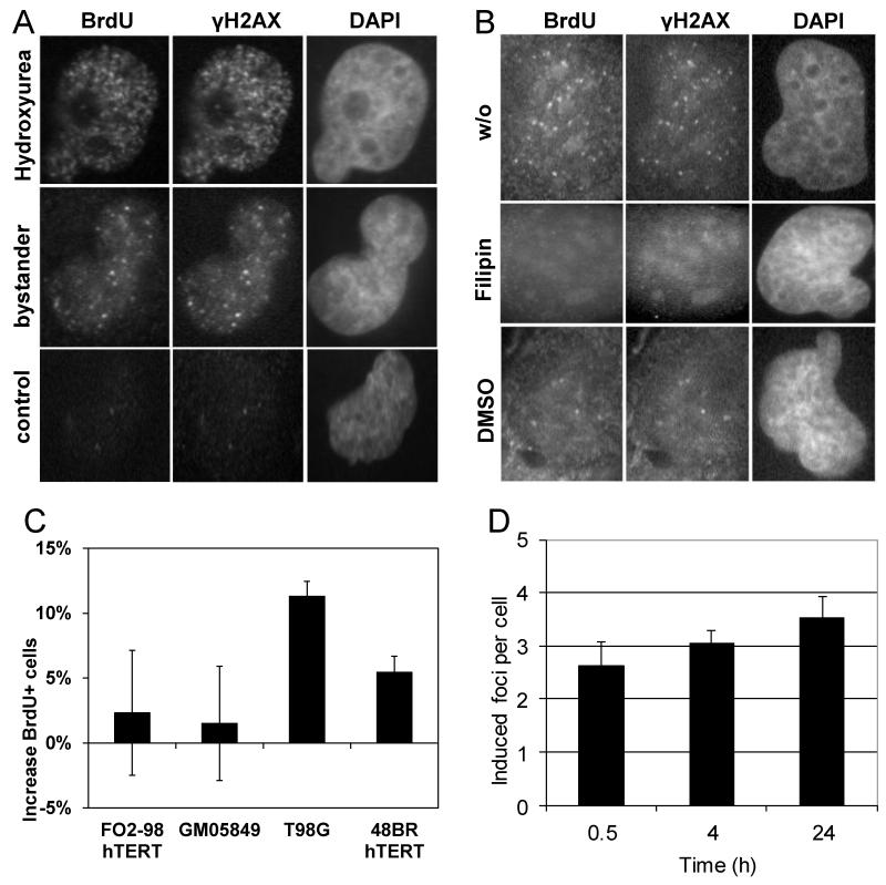
Immunofluorescence microscopic visualisation of stalled replication forks in bystander T98G cells. (A) Co-localisation of BrdU foci with γH2AX nuclear foci at sites of stalled replication in bystander cells and cells treated with 20 μM hydroxyurea; (B) Inhibition of BrdU and γH2AX nuclear foci in bystander cells by Filipin and DMSO. (C) Increase in the fraction of BrdU-positive cells detected by flow cytometry following pulse-labelling of bystander and corresponding control cultures. Bars show average values from 3 independent experiments for each cell line. Error bars show the associated standard errors. (D) Persistent increase of bystander gamma-H2AX foci numbers in T98G cell cultures treated for 0.5, 4 or 24 h with conditioned medium. Bars show average values from 2-6 independent experiments for each time point. Error bars show the associated standard errors.

