
Figure 3. Circuit diagrams of ganglion cells not identified in the connectome.
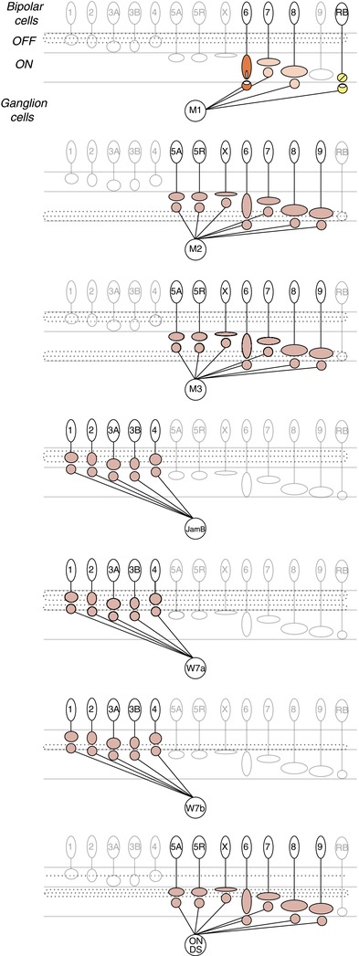
Ganglion cell types missing from the connectome include the melanopsin-expressing ganglion cells, M1, M2, M3; JamB; W7a; W7b; and the ON direction selective (ON DS) ganglion cells. Evidence from physiology and synaptic markers indicates inputs from ON cone bipolar cells to the M1 melanopsin ganglion cell (orange). En passant synapses from either the type 6, 7, or 8 ON cone bipolar cells have been speculated by Dumitrescu et al. (2009). Both the M2 and M3 melanopsin ganglion cells receive excitatory ON input (Schmidt & Kofuji, 2010; Schmidt et al. 2011a,b2011b); however, the bipolar cell type providing input remains unknown, hence we indicate a low level of certainty from physiology (red). The JamB, W7a and W7b ganglion cells described by Kim et al. (2010) depolarize to light decrements in their receptive field centres, suggesting that these cells may receive inputs from OFF cone bipolar cells. These synapses are coloured by the lowest level of certainty for the method of physiology (red). The majority of ON direction selective ganglion cell dendrites stratify in the ON sublamina of the inner plexiform layer and their membrane potential depolarizes to light increments, suggesting inputs from ON cone bipolar cells (Dhande et al. 2013). However, a particular type has not been implicated, thus the level of certainty for each type is low.