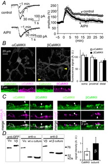
Figure 1. α- and βCaMKII in a PN.

A, representative traces and time courses of GABA current amplitudes before and after the conditioning depolarization in the absence (control) or presence of AIPII (1 μm). n = 5 for each. B, immunofluorescence images of endogenous α- or βCaMKII in PNs. The area of a PN was defined as the calbindin-positive area (not shown). Yellow arrowheads indicate βCaMKII expression in neurons other than a PN, which were calbindin-negative. Bottom, high-resolution images of each CaMKII subunit (magenta) and calbindin (green, a PN marker). Right, fluorescence intensities of anti-αCaMKII and anti-βCaMKII signal in soma, proximal and distal dendrites. Staining efficiency of each antibody is calibrated, and the signal is normalized to that of βCaMKII in soma. n = 20 cells for each. C, representative immunofluorescence images of GABAAR α1 or VGAT, and α- or βCaMKII. D, western blotting of Venus-tagged CaMKII subunits (Vα and Vβ) and wild-type CaMKII subunits (wt α and wt β) prepared from HEK293T cells, and endogenous α- and βCaMKII prepared from a cerebellar culture (n = 3 cultures). Quantified signals for α- and βCaMKII after calibration of staining efficiency of each antibody are shown. A.U., arbitrary units.
