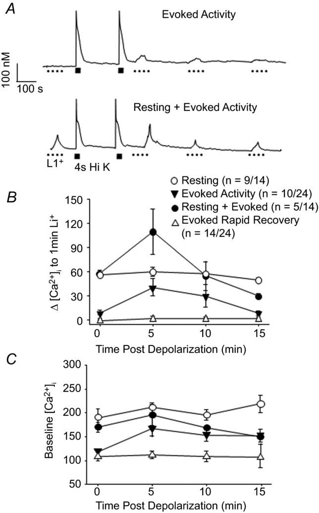
Figure 4. Characterization of ‘resting’ NCX activity in putative nociceptive cutaneous DRG neurons.

A, examples of neurons in which Ca2+ transients were evoked with Li+. In one neuron (top trace), Li+ (1 min) evoked transients were only detected after stimulating the neuron with high K+ and the amplitude of these transients decayed over time. In the second (bottom trace), Li+-evoked transients were evoked before and after stimulating the neuron with high K+. In this neuron, the amplitude of the Li+-evoked transient increased following high K+ stimulation, but decayed to baseline levels over time. B, the amplitude of the Li+-evoked transient in 4 types of putative nociceptive cutaneous DRG neurons are plotted as a function of time relative to high K+ stimulation as illustrated in A, where 0 is before and 15 indicates 15 min after high K+-induced depolarization. C, resting [Ca2+]i data for the four groups of neurons plotted in B, where resting [Ca2+]i was determined 10 s prior to Li+ application.
