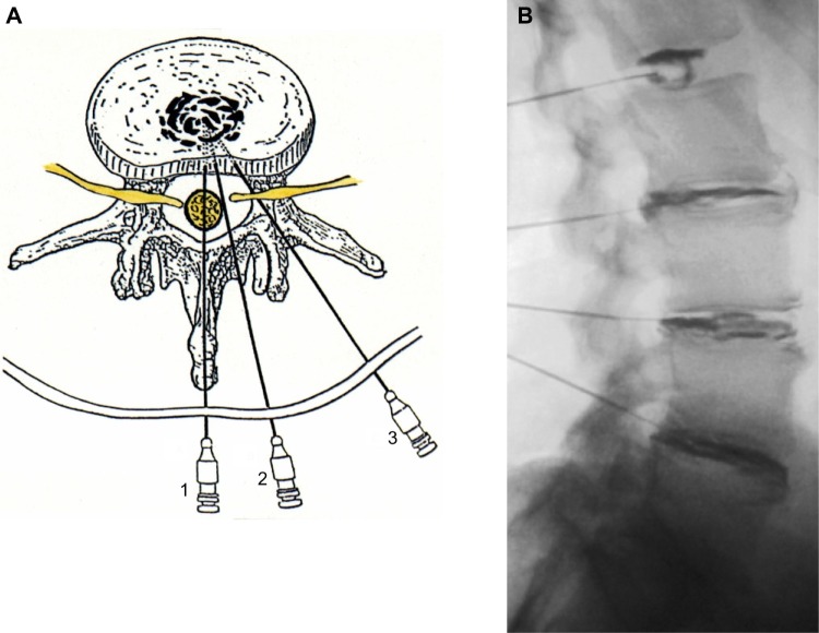
Figure 2.
(A) A schematic axial view of the intervertebral disk with preferred location of the needle tip in the center. Needle-approach 3 is recommended as approaches 1 and 2 have a higher risk of leakage of cerebrospinal fluid with subsequent postprocedural headache. (B) A radiographic image of the lumbar spine after multilevel diskography showing in the upper disk an intact annulus fibrosus (contained contrast agent in the nucleus pulposus). The lower 3 disks show degeneration with annular tears as evidenced by leakage of contrast agent to the outer disk.

