Fig. 2.
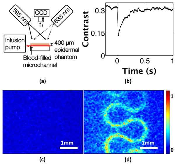
In vitro photothermal LSI of blood flowing through a microchannel. (a) Photothermal LSI setup with blood infused into the system at 4 mm/s and a 400 μm epidermal phantom placed above the microchannel. (b) Speckle contrast versus time in a region of interest above the channel shows a 60% decrease in flow occurring after the excitation pulse at time 0 s, and returning to baseline over the next ~0.4 s. (c) Normalized speckle flow index (SFI) in microchannel before the photothermal excitation (Media 1). (d) SFI image after the excitation pulse (Media 1) shows the blood flow in the channel clearly visible beneath the skin-simulating phantom.

