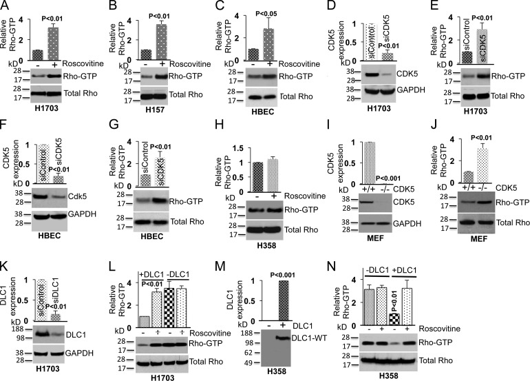
Figure 3.
CDK5 regulates RhoGTP levels through DLC1. (A) Treatment of a DLC1-positive H1703 line with Roscovitine (10 µM) increases RhoGTP (top) but not total Rho (bottom). The graph represents the relative RhoGTP ± SD from three independent experiments. (B and C) Experimental conditions were similar for H157, which is a second DLC1-positive NSCLC line (B), and for the HBEC cell line, which is an untransformed human lung epithelial cell line (C). (D) Knockdown of CDK5 by siRNA (top); GAPDH was used as a loading control (bottom). The graph represents the relative CDK5 expression ± SD from three independent experiments. (E) Knockdown of CDK5 increases RhoGTP (top) but not total Rho (bottom). The graph is as in D. (F and G) Knockdown of CDK5 expression in HBEC cells leads to an increase in RhoGTP. (H) Roscovitine does not alter RhoGTP in the DLC1-negative H358 NSCLC line. (I) Expression of CDK5 in MEFs cells isolated from WT and CDK5−/− embryos (top). GAPDH was used as loading control (bottom). (J) RhoGTP is higher in CDK5−/− MEFs than in WT MEFs (top). Total RhoGTP is similar in both MEFs (bottom). (K and L) Knockdown of DLC1 in H1703 renders the RhoGTP in the cells unresponsive to Roscovitine. (K) Knockdown of DLC1 expression (top). GAPDH was used as a loading control (bottom). (L) RhoGTP (top) and total Rho (bottom) in DLC1 siRNA– or control siRNA–transfected H1703 cells in the absence or presence of Roscovitine. Knockdown of DLC1 abrogates the ability of Roscovitine to affect the level of RhoGTP. (M and N) Transfection of DLC1 into DLC1-negative H358 cells enables RhoGTP to be regulated by CDK5. (M) Expression of DLC1-WT in stably transfected H358. (N) RhoGTP (top) and total Rho (bottom) in H358 cells, with or without transfected DLC1, incubated in the absence or presence of Roscovitine. Data in each panel are representative of three independent experiments. Error bars indicate ± SD.

