
Figure 6. Regulation in HuH-7 and HK-2 cells of WIP1 by ATO and effects on Cis-P cytotoxicity.
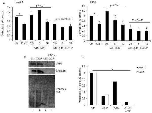
A. MTT cell viability assays in HuH-7 and HK-2 cells showed ATO was cytotoxic in both cell lines in 10 μM or 5 μM and 10 μM concentrations, respectively, p<0.05, asterisks, Chi-square tests. Moreover, ATO increased Cis-P cytotoxicity in HK-2 cells, asterisks, p<0.05, whereas that was not the case in HuH-7 cells. B. Western blot showing inhibition of WIP1 compared with control cells (top panel, lanes 1, 2) in ATO-treated HK-2 cells either with or without Cis-P (top panel, lanes 3, 4). Also, reprobing of the blot showed ATO decreased β-tubulin in these cells (middle panel, lanes 3, 4). Ponceau red staining after completion of westerns confirmed proteins were present in all samples (bottom panel). C. Analysis of SP cells after ATO and Cis-P in HuH-7 and HK-2 cells indicated these were depleted in both cases, asterisk, p<0.05. In HK-2 cells, SP fractions were no longer detectable.