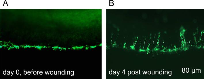
Figure 1.
Transduction of organ-cultured diabetic cornea with Ad-EGFP (live images). A, only limbal region is transduced (EGFP+ cells, green). B, during healing of a large 8.5 mm wound, limbal cell migrate centripetally. Days are relative to wound healing study. e, epithelium, s, stroma. Bar = 80 μm.

