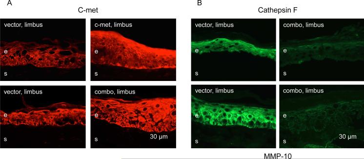
Figure 2.
Verification of viral transduction by immunostaining. A, limbal c-met expression is increased upon limbal transduction with Ad-c-met or combined Ads with MMP-10 and cathepsin F shRNA and full-length c-met (combo). B, both MMP-10 and cathepsin F expression in the limbus is markedly decreased upon combo treatment. Here and below, representative pictures were taken with the same exposure times for each row and each marker. Here and below, the transduced components are indicated in white on the panels, and the markers revealed by immunostaining of corneal sections are indicated in black above and below the respective panels. e, epithelium, s, stroma. Bar = 30 μm.

