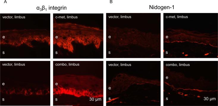
Figure 4.
Normalization of patterns of diabetic markers upon limbal gene therapy. A, epithelial α3β1 integrin immunostaining becomes markedly stronger and more regular in the limbus following c-met overexpression and combo treatment. B, staining for nidogen-1 in the limbal basement membrane after vector transduction is weak and discontinuous over large areas (arrows). It becomes bright and nearly continuous following both treatments. e, epithelium, s, stroma. Bar = 30 μm.

