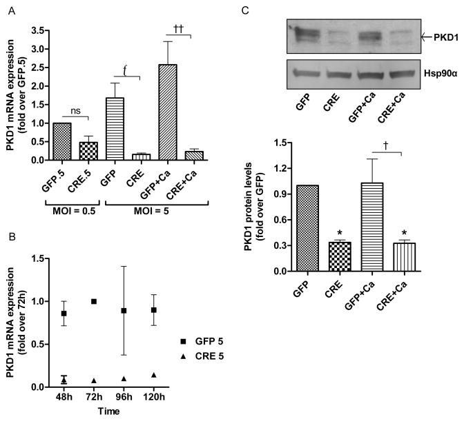
Fig 1. PKD1 depletion is achieved with adenoviral Cre-recombinase in floxed PKD1 keratinocytes.
(A–C) Keratinocytes from floxed PKD1 neonatal mice were infected with Cre-recombinase (CRE)- or GFP vector (GFP)-expressing adenoviruses for 24h. The medium was replaced with 50μM calcium-containing control medium or elevated calcium (125μM calcium; Ca)-containing medium at 48h (post-infection) for 24h, and then cells were harvested for qRT-PCR or Western analysis. (A–B) Quantitative RT-PCR was performed to determine the expression of the PKD1 gene (A) for the two indicated multiplicities of infection (MOI) and (B) for different time points of infection. (C) Western blot (upper panel) and densitometric analysis of multiple Western blots (lower panel) of PKD1 protein, with Hsp90α as the loading control, are shown. Please note that based on previous studies, the middle band represents unphosphorylated PKD1 as indicated by the arrow, the lower band represents PKD2 and the upper band(s) represent phosphorylated, activated PKD1 and PKD2. Data represent the means ± SEM from at least 3 independent experiments.

