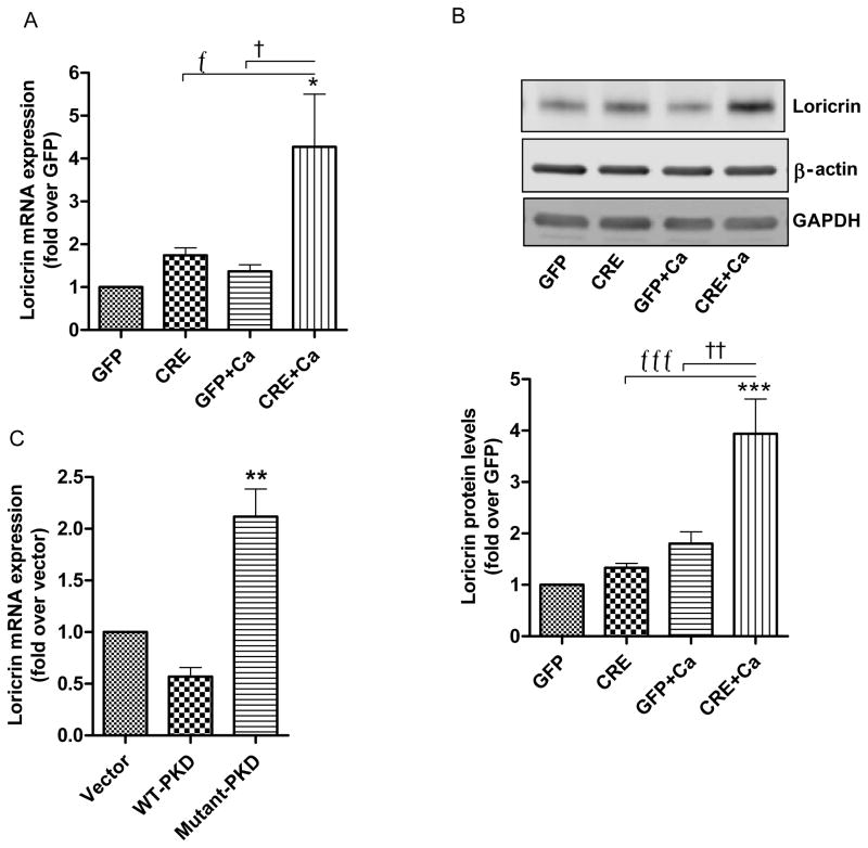
Fig 2. Effect of PKD1 depletion on a late differentiation marker, loricrin.
(A–B) Keratinocytes from floxed PKD1 neonatal mice were infected with Cre-recombinase (CRE)- or GFP vector (GFP)-expressing adenoviruses for 24h. The medium was replaced with 50μM calcium-containing control medium or elevated calcium (125μM calcium; Ca)-containing medium at 48h (post-infection) for 24h. (A) Quantitative RT-PCR was performed to determine the expression of the loricrin gene. (B) A representative Western blot (upper panel) and a densitometric analysis (N=4) (lower panel) of loricrin protein, with β-actin and GAPDH as loading controls, are shown. (C) Sub-confluent keratinocytes from CD-1 neonates were infected with the indicated adenoviral constructs for 24h and analyzed for loricrin mRNA expression using qRT-PCR. Data represent the means ± SEM from at least 3 independent experiments.

