Figure 3.
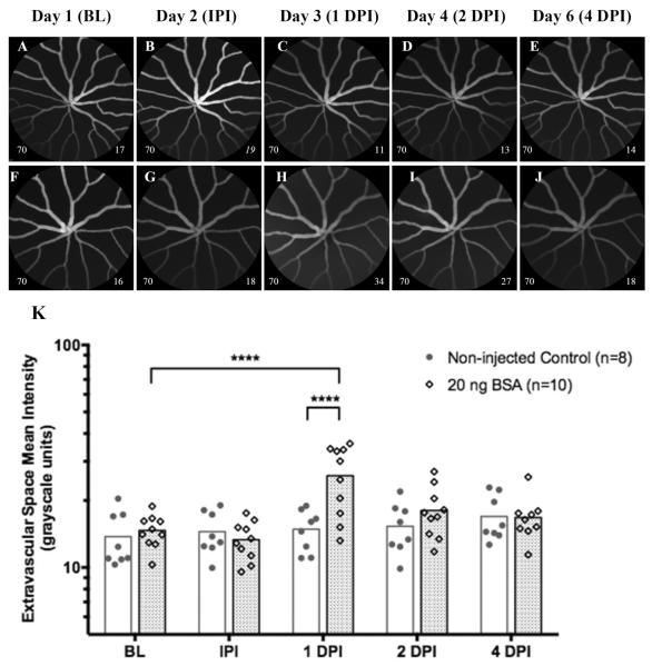
Representative images (Fig. 3A–J) and graphical results (Fig. 3K) from an experiment involving IOI of BSA (20 ng, 0.8μl) in a group of Tg(l-fabp:DBP-EGFP) zebrafish compared to another group that did not receive injections. Fig. 3A–E and 3F–J show representative images collected from a non-injected and BSA-injected animal, respectively. Repeated imaging sessions in the injection-free group did not significantly change avascular space mean signal magnitude over time (Fig. 3K). In contrast, animals that received injections of BSA had a significant increase in mean signal magnitude at 1 DPI and can be observed in Fig. 3H as extravascular haze. This increase was significant relative to both the original mean baseline level on day 1, as well to the mean level observed at 1 DPI for the non-injected group. This response was brief and resolved within 24 hrs to an insignificant level at 2 DPI. Important to note is that no increase in ASMI was observed IPI, which demonstrates that the IOI does not cause acute damage resulting in leakage of EGFP-albumin. Data shown as a scatter plot with bars to indicate group time point means. (Image notations: Lower left # − camera sensitivity setting; Lower Right # - ESMI; normal style text- ESMI obtained with sensitivity set @ 70, italicized text- ESMI was corrected).

