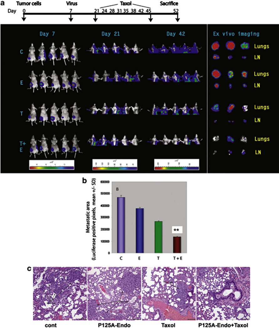
Fig. 3. Effect of antiangiogenic gene therapy and paclitaxel on metastasis of orthotopically transplanted human breast cancer cells (intervention model).
MDA-MB-231-luc cells were transplanted into mammary fat pads of female, athymic mice. Whole body images were obtained on a weekly basis after intraperitoneally administration of luciferin in a Xenogen system (a). Luminescence signals from the primary tumors were too strong and were overlapping with the signals originating from the metastatic sites. Therefore, the primary tumors were shielded (lower half) to reveal tumor metastasis to lungs and lymph nodes (days 21 and 42). Intensity of signals is shown at the bottom. On day 42, mice were killed and brachial lymph nodes and lung tissues were removed for ex vivo imaging. C, adeno-associated virus (AAV)-LacZ; E, AAV-P125A-endostatin; T, paclitaxel; T+E, combination treatment; LN, lymph node. Panel (b) shows relative luciferase-positive pixels from the metastatic sites. Each value is a mean of five animals ± s.d. Panel (c) shows the histopathology of lungs. The lung tissues were stained with hematoxylin and eosin stain and the black arrow shows the tumor that has metastasized to the lungs (×200). **Denotes statistical significance (P<0.02). Reprinted with permission from ref 105.

