Figure 6.

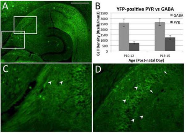
ChR2-YFP distribution in the hippocampus at age P14 in the Thy1-ChR2-YFP transgenic mouse. A. Transverse hippocampal section showing high expression in the subiculum, lower expression in the CA3/CA1 regions, and lack of expression in the dentate gyrus. Higher magnification of the areas indicated in A are shown in panels C and D. Arrowheads identify YFP-expressing cells that appear to be interneurons, while arrows (D only) indicate a minority of cells that appear to be YFP-positive pyramidal neurons. B. Comparison of the number of YFP-positive cells and for the PYR and GABA cell types across the age range used for in vitro studies. While the number of PYR cells expressing ChR2-YFP increased with age, the numbers of YFP positive PYR cells was still smaller compared with that of YFP positive GABA neurons. Similar expression pattern was found in mice even at age P100 that were used for in vivo experiments (data not shown). Scale bar, 500μm.
