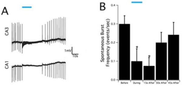
Figure 7.

Suppression of epileptiform activity by blue laser in the in-vitro hippocampal slice. A. Optical stimulation, indicated by blue bar, was applied at 20 Hz in the CA3 region of the hippocampal slice and the epileptiform activity was suppressed both in the CA1 and CA3 regions. B. The summary of spontaneous burst frequency in the CA3 region before, during, and after optical stimulation. Optical stimulation could cause significant reduction during and 15s after stimulation (p<0.05) and then the burst frequency gradually return to the baseline level.
