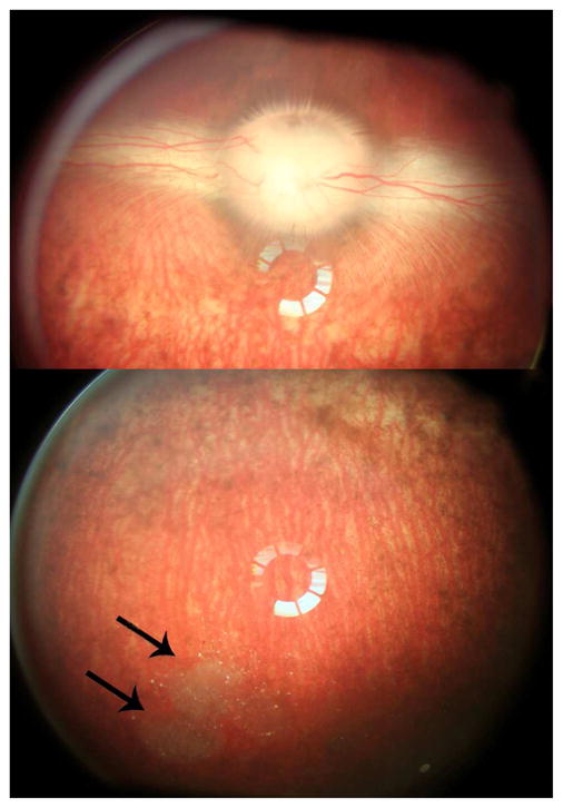
Figure 9.

Color fundus photograms taken on day 14 before sacrifice. The upper panel photo demonstrated normal clear media, normal optic nerve head and medullary ray. The lower panel photo showed clear media, normal inferior retina, as well as the depot of cov-OPS-COO-DEX particles away from visual axis and at the inferior vitreous cavity (arrows). The rings at the centers of the images were from a light reflex from the pre-place 20 diopter lens in the front of the camera lens for wider viewing purpose.
