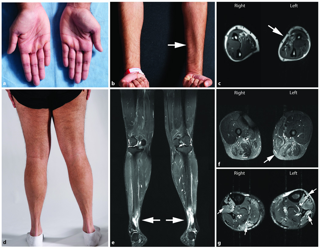
Figure 2.
Physical and MRI findings in a patient with GNE myopathy presenting with left hand weakness. (a) Photograph of hands showing atrophy of the hypothenar area, more pronounced on the left. (b) Photograph and (c) T1-weighted axial muscle MRI of the forearms show atrophy of forearm flexor muscles, more prominent on the left arm (arrows). Involvement of the distal lower extremities is evident on (d) a photograph of posterior lower extremities and (e) the T2-weighted STIR coronal muscle MRI image of the lower extremities.
T2-weighted STIR muscle MRI images of the lower extremities including coronal (e) and axial views of the thigh (f), and lower leg (g), show T2 hyperintense areas (arrows) noted bilaterally within the anterior tibialis muscles (e and g) gastrocnemius, flexor hallux longus and the soleus muscles (g), and patchy T2 signal hyperintense areas on the posterior thigh involving the adductor and hamstring muscle groups bilaterally (f).

