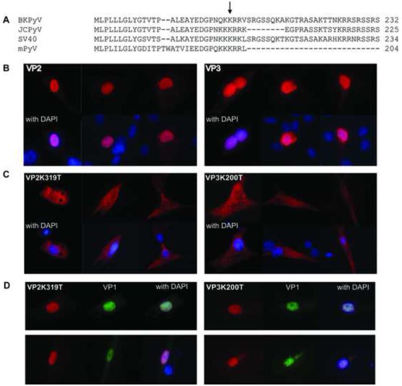
Figure 1. Subcellular localization of NLS mutant VP2 and VP3 proteins.
(A) VP3 Lysine 200 is conserved in a number of polyomavirus species. (B) Wild type VP2 and VP3 expression plasmids were transfected in RPTE cells, fixed at 24 h, and stained with anti-VP2/3 (red). Nuclei were stained with DAPI (blue). Both VP2 and VP3 are nuclear, as demonstrated by co-staining (bright purple) with DAPI. (C) RPTE cells were transfected with mutant VP3 (VP3K200R) or VP2 (VP2K319R) in the same manner. (D) RPTE cells were co-transfected with VP1 and either VP2 or VP3, and stained for VP2/3, VP1, and DAPI.

