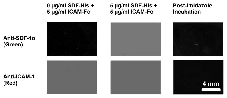
Figure 3. Characterization of uniformly coated protein surfaces by fluorescent antibody binding.

Fluorescent antibody binding to the surface was specific; insignificant monoclonal anti-SDF-1 antibody binding (green) was seen for a surface contacted with 0 μg/ml SDF-His (Column 1, Row 1), whereas significant antibody binding (green) was observed on surfaces contacted with 5 μg/ml SDF-His (Column 2, Row 1). Likewise, fluorescent antibody binding to the surface was reproducible; significant and comparable monoclonal anti-ICAM-1 antibody binding (red) was seen on different substrates contacted with the same 5 μg/ml ICAM-Fc concentration (Column 1, Row 2; and Column 2, Row 2). Finally, Significant abolishment of fluorescence (Column 3) upon Imidazole incubation of fluorescent antibody bound protein immobilized substrates (5 μg/ml ICAM-Fc + 5 μg/ml SDF-His contacted) demonstrated that the binding of His-tagged SDF-1α to the nickel coated surfaces was mostly specific, implying correct orientation of the protein with active site accessible.
