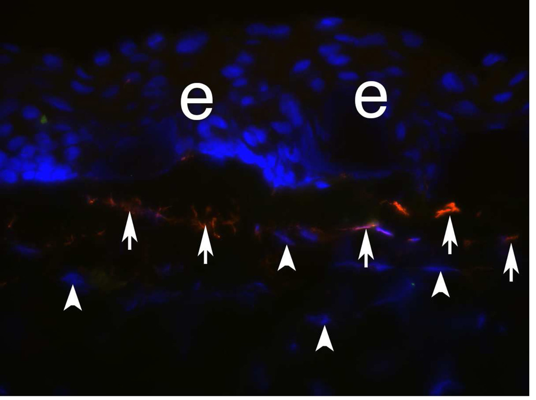
Fig. 5.
Vimentin+ cells (arrows) in the anterior stroma at one week after −9D PRK in a rabbit. e indicates epithelium. This is a rabbit cornea one week after −9D PRK that underwent double immunohistochemistry for vimentin (orange) and α-SMA (green). At this time point after surgery, none of the cells in the anterior stroma express α-SMA. However, many of these vimentin+ cells are likely myofibroblasts in early development that will begin to express α-SMA with further maturation at about two weeks after surgery. Note keratocytes (arrowheads) that have been shown in prior studies to also express vimentin, but at much lower levels, and this expression was not detected with the concentration of primary antibody for vimentin used in this staining (see Chaurasia et al., 2009). Thus, these early myofibroblasts that are vimentin+SMA-express vimentin at high levels. 400X

