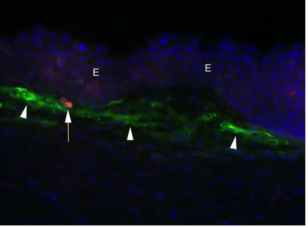
Fig. 8.
Apoptosis of myofibroblasts at one month after −9D PRK. TUNEL assay was used to detect apoptosis and immunohistochemistry to detect α-SMA in myofibroblasts (arrowheads) revealed one myofibroblast undergoing apoptosis (arrow). E indicates epithelium. Blue stain is DAPI for cell nuclei. 400X. The balance between myofibroblast generation and myofibroblast apoptosis in a particular cornea after injury determines whether haze is increasing, persisting, or disappearing over time. After Wilson, Chaurasia, and Medeiros, F.W. 2007 with permission.

