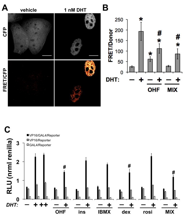
Figure 6.
Human adipocyte differentiation alters the conformation of AR. (A) HeLa cells transiently transfected with CFP-AR-YFP were treated with vehicle, 10 uM OHF, or differentiation media (MIX) in the presence or absence of 1 nM DHT for 20 h. Scale bar, 10 μm. (B) FRET imaging was used to determine AR conformation (n≥10 cells/condition). (C) An N-terminal region containing AR 1-660 was fused to the VP16 TAD while a C-terminal region containing AR 624-919 (wild-type) was fused to the to the Gal4-DBD domain. HeLa cells were co-transfected with AR fragments and reporter constructs as described in Materials and Methods. After 12 h of transfection, cells were trypsinized and equally seeded into 96-well plates. Cells were allowed to re-attach and subsequently treated with dexamethasone (500 nM), IBMX (250 uM), insulin (200 nM), rosiglitazone (3 uM), MIX, AR ligands, alone or in the presence of 0.1 nM DHT. After 24 h of treatment, luciferase activity was detected. Data represents the units of firefly luciferase corrected for the units of renilla luciferase detected in the same plate. Shown is one representative experiment from 4 replications. p<0.05 was considered significant for treatments compared to vehicle (#) or DHT (*) treatments.

