
Figure 6.
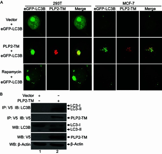
PLP2-TM colocalizes and coimmunoprecipitates with autophagy marker protein LC3. (A) HEK293T and MCF-7 cells were transfectd with pcDNA3.1 empty vector, PLP2-TM-V5. After 48 h post-transfection, the cells were fixed and incubated with anti-V5-tagged primary antibody, followed by being stained with an Alexa Fluor 594-conjugated goat anti-rabbit secondary antibody. Colocalization of PLP2-TM-V5 (red) and eGFP-LC3B (green) were observed using a confocal microscope as described in Fig. 1B. (B) HEK293T cells were transfected with pcDNA3.1 empty vector or PLP2-TM-V5 for 48 h, and the cell lysate was immunoprecipitated (IP) with an anti-V5 antibody and immunoblotted (IB) with an anti-LC3 and anti-V5-tagged antibody to detect expression of LC3 (top panel) and V5-tagged PLP2-TM (second panel), respectively. The whole cell lysate (WCL) was blotted with indicated antibodies to evaluate expression of endogenous LC3 and V5-tagged PLP2-TM. Beta-actin was analyzed to serve as a protein loading control