Figure 3.
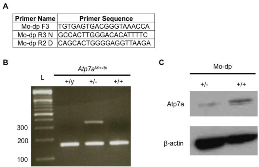
Genotyping and Atp7a expression in Mo-dp+/− mouse brains. A. Primers used to amplify normal and dappled alleles. The normal allele was amplified by the primers Mo-dp F3 and Mo-dp R3 N, producing a 167 bp amplicon. The Mo-dp allele was amplified by the primers Mo-dp F3 and Mo-dp R2 D yielding to a 354 bp amplicon. B. Duplex PCR to genotype and identify normal (167 bp band) and mo-dappled (354 bp band) alleles. All three viable genotypes are represented in this figure. C. Western blot analysis of total proteins from mouse brains shows lower Atp7a levels in the heterozygous female (Atp7aMo-dp+/−) compared to a normal female (Atp7aMo-dp+/+).

