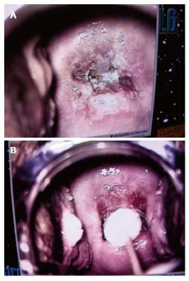
Figure 4.

Conization of human papilloma virus cervical intraepithelial neoplasia 3 lesions. Colposcopy of papillomavirus cervical intraepithelial neoplasia 3 lesions from a patient treated with conization. Photographs of cervix before (A) and immediately after conization (B).
