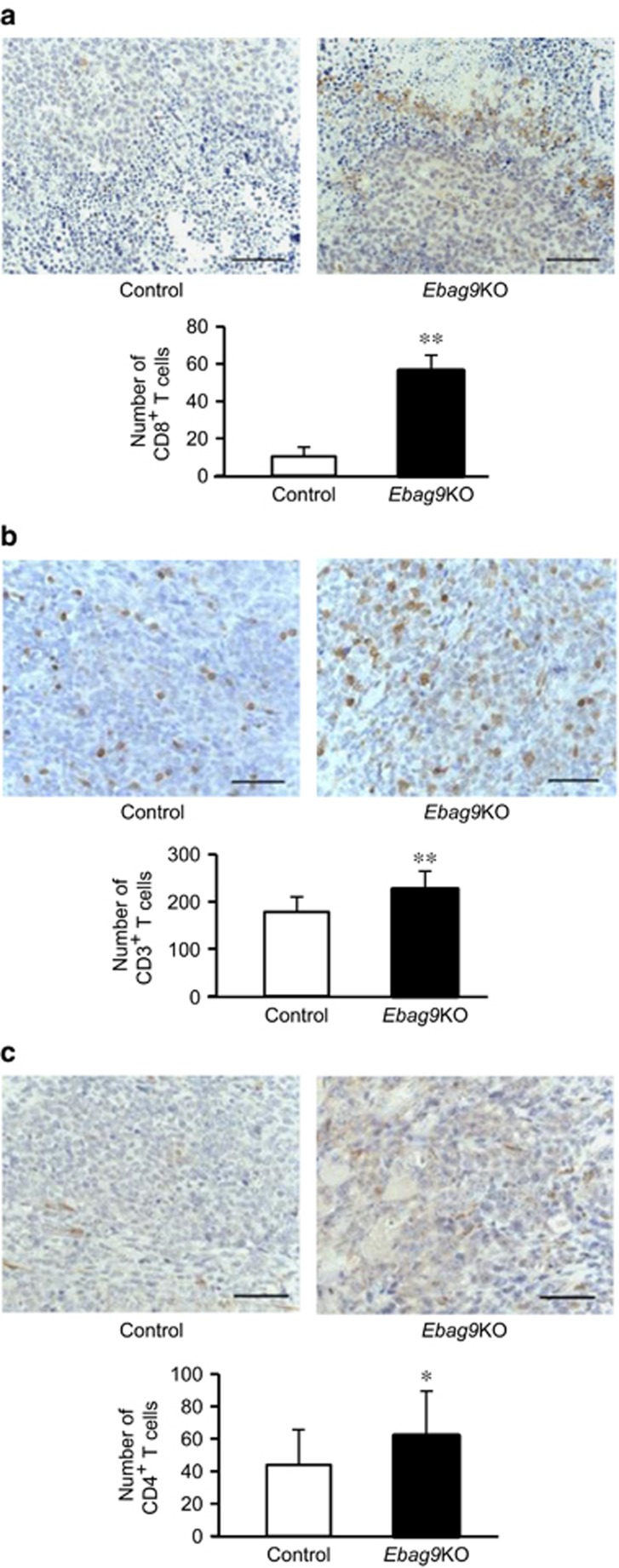
Figure 4.
Increased numbers of CD8+, CD3+ and CD4+ T cells in tumors generated in Ebag9KO mice. Paraffin sections of tumors generated in Ebag9KO and control mice were blocked in 0.3% H2O2 and incubated with specific antibodies for CD8 (a), CD3 (b) or CD4 (c). Sections were then incubated with antirabbit EnVision+ reagent and counterstained with hematoxylin. Tumor-infiltrating lymphocytes positive for CD8 (a), CD3 (b) or CD4 (c) expression were microscopically counted in a high-power field of view at a magnification of 400 × (Ebag9KO, n=8; control, n=6). The data shown in lower panels are mean values±s.d. *P<0.05; **P<0.01 by Student's t-test. Scale bar, 20 μm.

