
Figure 2.
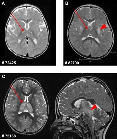
Brain MRI of Affected Individuals #72425, #82790, and #75168
(A) MRI of individual #72425 shows small T2 hyperintensities in the anterior thalamus bilaterally (arrow).
(B) In individual #82790, T2-weighted MRI shows bilateral hyperintensities in the putamen (arrowhead) and weakly also in the anterior thalamus (arrow).
(C) T2-weighted MRI of individual #75168 shows pronounced bilateral hyperintensities affecting the whole thalamus (arrow, axial view at the left) and extending to the mesencephalon (arrowhead, sagittal view at the right).