Figure 3.
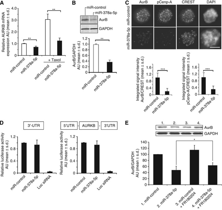
Excess of miR-378a-5p indirectly suppresses Aurora B during mitosis. Aurora B kinase mRNA (A) and protein (B) levels are significantly reduced by miR-378a-5p overexpression in HeLa cells (data are mean±s.d. from three replicate assays). (C) Representative micrographs showing loss of Cenp-A phosphorylation (Ser7) and Aurora B immunofluorescence signals in taxol-arrested miR-378a-5p-overexpressing cells in comparison with controls. CREST marks the centromeres. The graphs show quantification of the pCenp-A and Aurora B centromere signals normalised against CREST. The data are mean±s.d. from 30 cells, 20 centromeres quantified per cell. (D) The graphs show quantification of luciferase reporter assays indicating that miR-378a-5p does not bind to the 3′UTR or elsewhere of the Aurora B mRNA. Luciferase silencing was used as a positive control. Data are mean±s.d. from 4 to 5 replicate assays. (E) The Western blot and graph shows partial recovery of Aurora B protein levels in FR180204-treated HeLa cells with excess of miR-378a-5p in comparison with controls. Data are mean±s.d. from 3 to 5 replicate assays. The asterisks denote statistical significances (*P<0.05, **P<0.01, ***P<0.001). The scale bar (C)=10 μm. Abbreviation: AU=arbitrary units.

