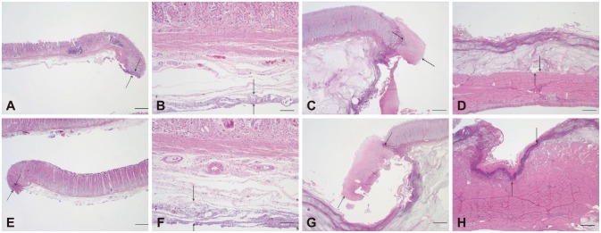
Fig. 5.

Representative histological findings of endoscopic mucosal dissection (ESD) specimens and residual muscularis propria tissue from the Optimos knife group (A-D) and combination knives group (E-H; H&E stain). (A, B) ESD showing minimal injury to the lamina propria and submucosa in the Optimos knife group. (C, D) Residual bed tissue showing minimal injury to the lamina propria and muscularis propria in the Optimos knife group. (E, F) ESD showing minimal injury to the lamina propria and submucosa in the combination knives group. (G, H) Residual bed tissue showing marked injury in the lamina propria and muscularis propria in the combination knives group. Arrows represent the extent of injury. Scale bars represent 50 µm in (B) and (F) and 500 µm in the others.
