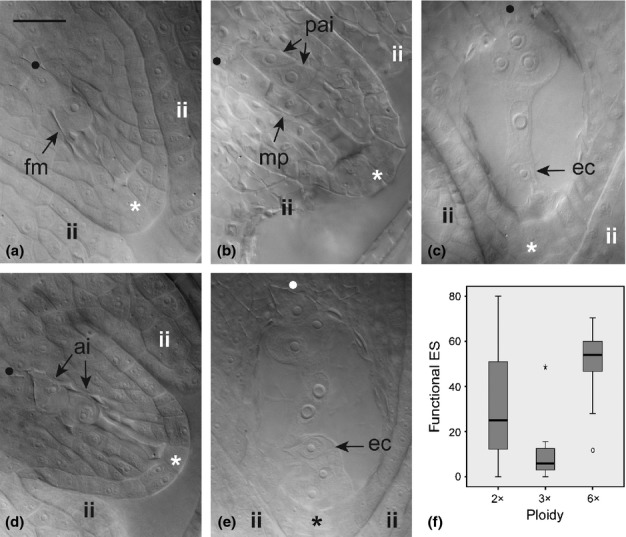
Fig 1.

Key female gametophyte reproductive stages during flower development in 2x homoploid and 3x heteroploid synthetic hybrids (a–c), and natural 6x heteroploid hybrids (d, e) of Ranunculus. Ovule images are placed as in Supporting Information Fig. S1 so that rotation angles can be tracked by the orientation of the chalazal-micropylar axis. (a) Functional megaspore immediately after meiosis; (b) end of meiosis showing four meiotic products – the two located toward the micropyle are aborted whereas the other two show signs of abortion – and two putative aposporous initial cells; (c) completely rotated ovule showing a mature seven-celled embryo sac at blooming stage; (d) aborted germline and two aposporous initials in the chalazal area; (e) mature embryo sac just before polar nuclei fusion to form a secondary nucleus in the central cell. (f) Box-and-whisker diagram for the proportion of functional embryo sacs (ES) among ploidy levels of interspecific hybrids. Boxes represent first and third quartiles, and the band inside each box indicates the median. Whiskers correspond to 95% CI. Outliers and extreme values are represented by circles and stars, respectively. Genotypes: (a) J5; (b) J20; (c) G13; (d) HöC29; (e) HöC35. fm, functional megaspore; mp, row of four meiotic products; pai, putative aposporous initial cells; ec, egg cell; ai, aposporous initial cells; ii, inner integuments; •, chalazal pole; *, micropylar pole. Bar, 30 μm.
