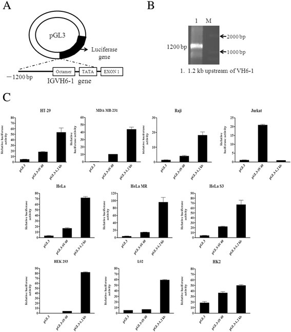
Figure 1.

The 5′-flanking sequence of VH6-1 exhibits promoter activity in non-B cancer cells. (A) Schematic diagram of 5′-flanking 1.2-kb pGL3 construct. (B) The 1.2-kb fragments amplified from upstream of VH6-1 in HT-29 cells by PCR. (C) The 1.2-kb pGL3 construct was transfected into HeLa, HeLa MR, HeLa S3, HT-29, MDA-MB-231, HEK 293,L02, HK2, Raji or Jurkat cells. Luciferase activity was measured using a dual-luciferase reporter system. The results are representative of three independent experiments after normalization to renilla luciferase activity (internal controls). Each bar represents mean ± SD.
