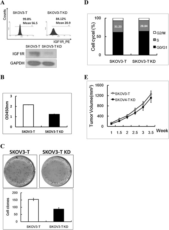
Figure 3.

IGF-1R knockdown could inhibit the proliferation of SKOV3-T. (A) IGF-1R expression was knocked down in SKOV3-T cells using lentivirus system and cells were analyzed by flow cytometry (up panel) and western blot (down panel) analysis; Cell proliferation (B) and clone formation (C) analysis both indicated that in SKOV3 KD cells, the cell growth was inhibited, while according to cell cycle analysis (D), SKOV3-T KD cells owned less S-phase cells in order to slower cell multiplification; In in vivo carcinogenic model, SKOV3-T KD exhibited slower tumor growth rate contrasting to SKOV3-T (E), indicating that IGF-1R was important to SKVO3-T cells when HER2-related cascade was blocked. To be clear, in this figure, “SKOV3-T” sample means control virus treated SKOV3-T cells.
