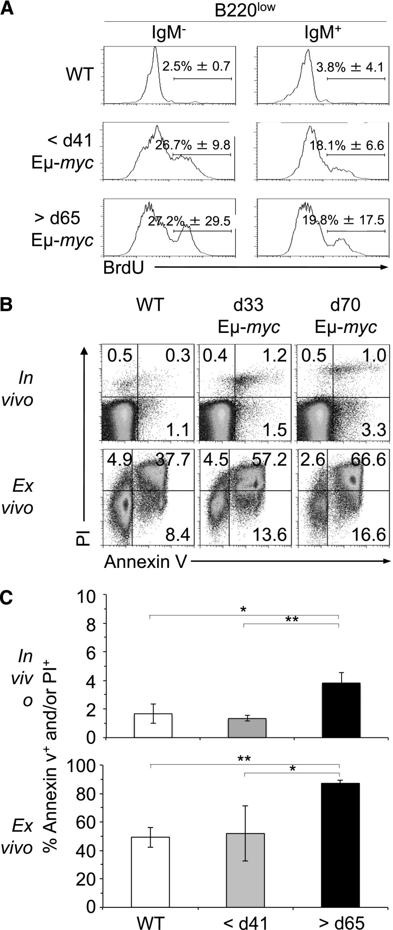
Figure 3.
Regression is not caused by changes in the rate of proliferation of tumor cells. (A) B220low cells of Eμ-myc and WT mice were stained for IgM and examined for BrdU incorporation and DNA content by flow cytometry. Eμ-myc mice at 33 and 70 ± 2 days of age and WT mice were injected intraperitoneally with 2 mg BrdU, and B220low blood cells were analyzed for IgM expression and BrdU incorporation 18 hours later. Numbers in quadrants indicate mean percentage of BrdU+ cells ±SD in each. Dot plots are representative of 3 independent experiments. (B-C) Detection of apoptotic cells by PI and annexin V staining of B220low cells from blood of WT mice at 33 days of age and Eμ-myc mice at 33 and 70 ± 2 days of age (>5 n each; upper panels). B220low cells of WT and Eμ-myc mice were also cultured for 5 hours before staining with PI and annexin V (>5 n each; lower panels). PI- and/or annexin V–positive cells were considered apoptotic. P values are indicated as HP < .05 and HHP < .01. d, day.

