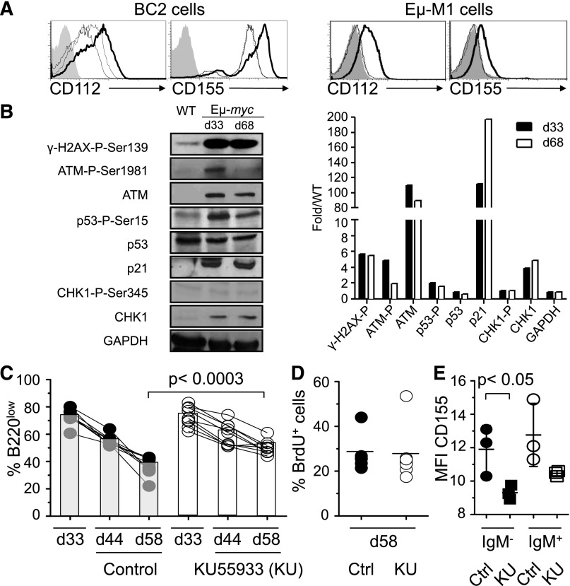
Figure 6.
Regression of B220low tumor cells in blood depends on the DNA damage response. (A) BC2 or Eμ-M1, cell lines derived from Eμ-myc mice, were treated with DMSO (fine line) or 10 μM Ara-C (bold line), a DNA-damaging agent, for 16 hours and stained for CD112 and CD155 expression. Some cells were pretreated with the 7.7 mM of the ATM/ATR inhibiting drug caffeine for 1 hour before treatment with Ara-C (dashed line) or DMSO (dotted line) for 16 hours. Filled histogram represents Ara-C–treated cells stained with isotype control. (B) Immunoblot analysis of purified B220low cells (>98% purity) from Eμ-myc mice at 33 and 68 ± 2 days of age or WT mice probed with antibodies for indicated DNA damage response markers. Levels were quantitated and normalized to WT levels (right panel). Shown is 1 out of 3 representative experiments. (C-E) Eμ-myc mice were injected intraperitoneally with 5 mg/kg KU55933 (n = 9) or vehicle (n = 8) at 37, 39, 47, 49, and 54 days of age. Percentage of B220low cells in the blood of an individual Eμ-myc mouse was determined at 33, 44, and 58 days of age by flow cytometry (C). KU55933-treated (open circles) or vehicle-treated (filled circles) mice also received 1 mg BrdU at 57 days of age. B220low tumor cells were stained for incorporation of BrdU and the percentage of BrdU+ B220low tumor cells was determined by flow cytometry at 58 days of age (D). Thirty hours after first injection of KU55933 (squares) or vehicle (circles), B220low cells were stained for CD155 and IgM expression. Mean fluorescence intensity of CD155 expression on IgM−B220low (filled symbols) and IgM+B220low (open symbols) cells is depicted (E). Statistical significance is indicated. Ctrl, control; d, day; MFI, mean fluorescence intensity.

