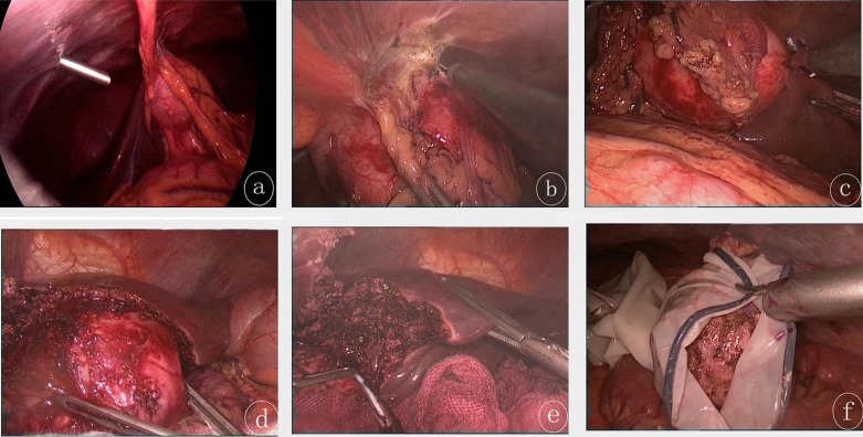
Figure 1.
(a) The echinococcosis cyst was located in the left lobe of the liver as revealed by laparoscopy. The cyst was adjacent to the peripheral tissues. (b) Monopolar electrocoagulation was performed to free the cyst from the peripheral tissue. (c) The cyst was separated from the liver using an ultrasound scalpel. (d) The ultrasound scalpel was used to dissociate the cyst and the liver adventitia, by delineating the hepatic interspace between the cyst and the liver. (e) The remaining liver wound after removal of the cyst. (f) The cyst was placed in a surgical bag designed by our team, using a latex medical glove: the front part (also called finger part) of the glove was cut using scissors, and served as the entry to the bag. The opening of the glove was ligated using a suture. The cyst was then removed from the abdomen of the patient.

