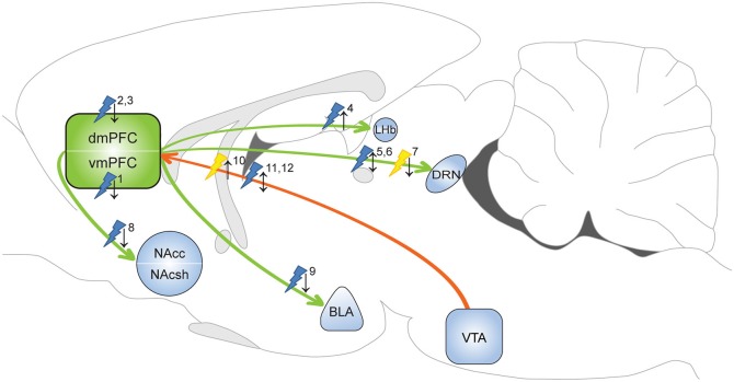
Figure 1.
Optogenetic evidence for the involvement of the mPFC in depressive-like behavior and anxiety. Yellow flash: photoinhibition; blue flash: photoactivation; ↑ = pro-depressive/anxiogenic effects; ↓ = antidepressant/anxiolytic effects. 1Covington et al. (2010): photoactivation increased sucrose preference and restored social interaction in defeat-susceptible mice. 2Kumar et al. (2013): photoactivation layer V pyramidal cells decreased immobility FST in naïve animals. 3Kumar et al. (2013): photoactivation layer V pyramidal cells increased time in open arms EPM test in defeated animals. 4Warden et al. (2012): photoactivation of mPFC-LHb projection promoted immobility FST in naïve animals. 5Warden et al. (2012): photoactivation of mPFC-DRN projection decreased immobility FST in naïve animals. 6Challis et al. (2014): photoactivation of vmPFC-DRN projection reduced social interaction in naïve animals. 7Challis et al. (2014): photoinhibition of vmPFC-DRN projection prevented social withdrawal in defeated animals. 8Vialou et al. (2014): photoactivation of dmPFC-Nac projection prevented social withdrawal. 9Vialou et al. (2014): photoactivation of dmPFC-BLA projection increased time in open arms EPM test. 10Chaudhury et al. (2013): photoinhibition of VTA-mPFC DA projection reduced social interaction in sub-threshold defeat animals. 11Friedman et al. (2014): photoactivation of VTA-mPFC DA projection restored social interaction in defeat-susceptible mice. 12Gunaydin et al. (2014): photoactivation of VTA-mPFC DA projection evoked anxiety-like behavior and place avoidance in naïve mice. dmPFC: dorsal medial prefrontal cortex; vmPFC: ventral medial prefrontal cortex; NAcc: nucleus accumbens core; NAcsh: nucleus accumbens shell; LHb: lateral habenula; DRN: dorsal raphe nucleus; BLA: basolateral amygdala; VTA: ventral tegmental area.

