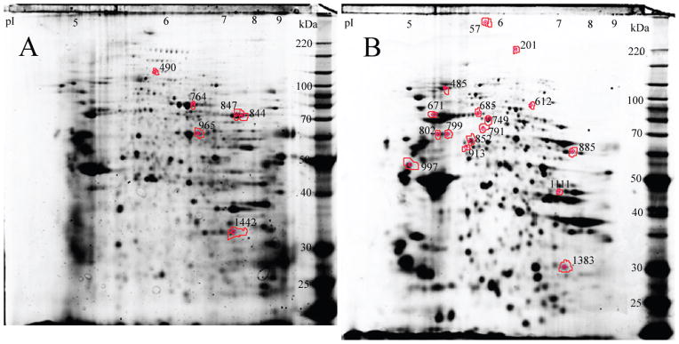
Figure 3.

Monocyte signatures of sickle cell crisis rate. Position of monocyte protein spots separated on a 2D IEF-SDS PAGE gel stained with Sypro Ruby that were successfully identified by tandem mass spectrometry. Monocyte proteins from the membrane fraction (Panel A) and cytosolic fraction (Panel B).
