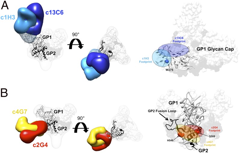
Fig. 3.
Details of the glycan cap and GP1-GP2 interface epitopes. (A) Competition analysis indicated that antibodies c13C6 and c1H3 have overlapping epitopes. Here, structures of c13C6 (dark blue) and c1H3 (light blue) bound to GPΔTM are illustrated, with the GPΔmuc crystal structure (PDB ID code 3CSY) fit into the GP EM density. GP1 is white and GP2 is black. Superimposition of the structures illustrates that the antibodies have overlapping epitopes within the glycan cap on GP1 (side view on the far left, top view in the center and far right). The footprints of these antibodies are highlighted on the far right. The mesh portion of the reconstruction is the part of the GP glycan cap that is resolved in the c1H3:GPΔTM structure. (B) As in A but c4G7 is in yellow and c2G4 is in red (side view on the far left and right, top view in the center).

