Fig. 2.
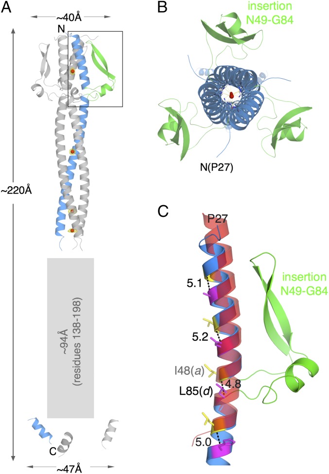
The crystal structure of NadA5. (A) Cartoon diagram of trimeric NadA5. Monomeric NadA5 that occupies the asymmetric unit is colored in blue for the coiled-coil region and in green for the wing-like insertion of the head; two symmetry-related molecules are shown in gray. Red spheres show the positions of iodide ions buried in the coiled-coil, and yellow meshes around these spheres show anomalous difference electron densities. The large rectangular gray-shaded outline shows the region of low-σ electron density. (B) Top view of NadA5, looking down the c axis, after rotation of 90° around x. (C) A zoomed-in view of the region of the insertion in the head of NadA5 (boxed region in A). The ribbon of NadA is colored as in A, and a superimposed α-helix, chosen from the trimeric transcription factor GCN4 (PDB ID code 4DME) to show the continuity of the coiled-coil of NadA5 around the region of the interruption, is shown as a semitransparent red ribbon. Yellow and magenta sticks depict NadA heptad residues a and d, respectively, and the distance in angstroms between their Cα carbons is shown.

