Fig. 1.
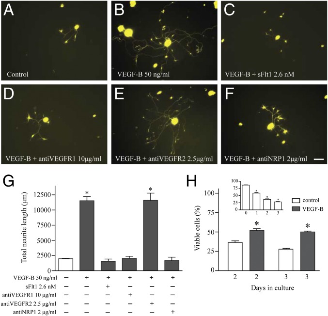
VEGF-B–induced neurite growth and survival on TG neurons via VEGFR1 and NRP1. Neurons from TG of thy1-YFP mice were isolated and cultured as indicated in Methods. (A) After 3 d in culture, few neurites grew in neurons cultured only in medium. (B) Addition of VEGF-B immediately after plating the cells induced long and extensive branching of neurites. (C) The neurite growth was competitively inhibited by soluble VEGFR1 at an equimolar dose or (D) by antibodies against VEGFR1 or (F) neuropilin 1, (E) but not by antibodies against VEGFR2. (G) Quantification analysis of neurite length demonstrated that VEGF-B–induced growth requires activation of VEGFR1 and NRP1. (H, Inset) The effect of VEGF-B on neuronal survival, analyzed as described in Methods, showed that there was a time-dependent cell death of TG neurons after 3 d in culture, and only 25% of neurons were found viable at this time point. (H) However, neuronal viability increased to 50% when cells were treated with a single dose of 50 ng/mL of VEGF-B. Data represent the mean ± SEM of three independent experiments, and a P value < 0.05 was considered statistically significant between treatments. (Scale bar, 50 μm.)

