Fig. 5.
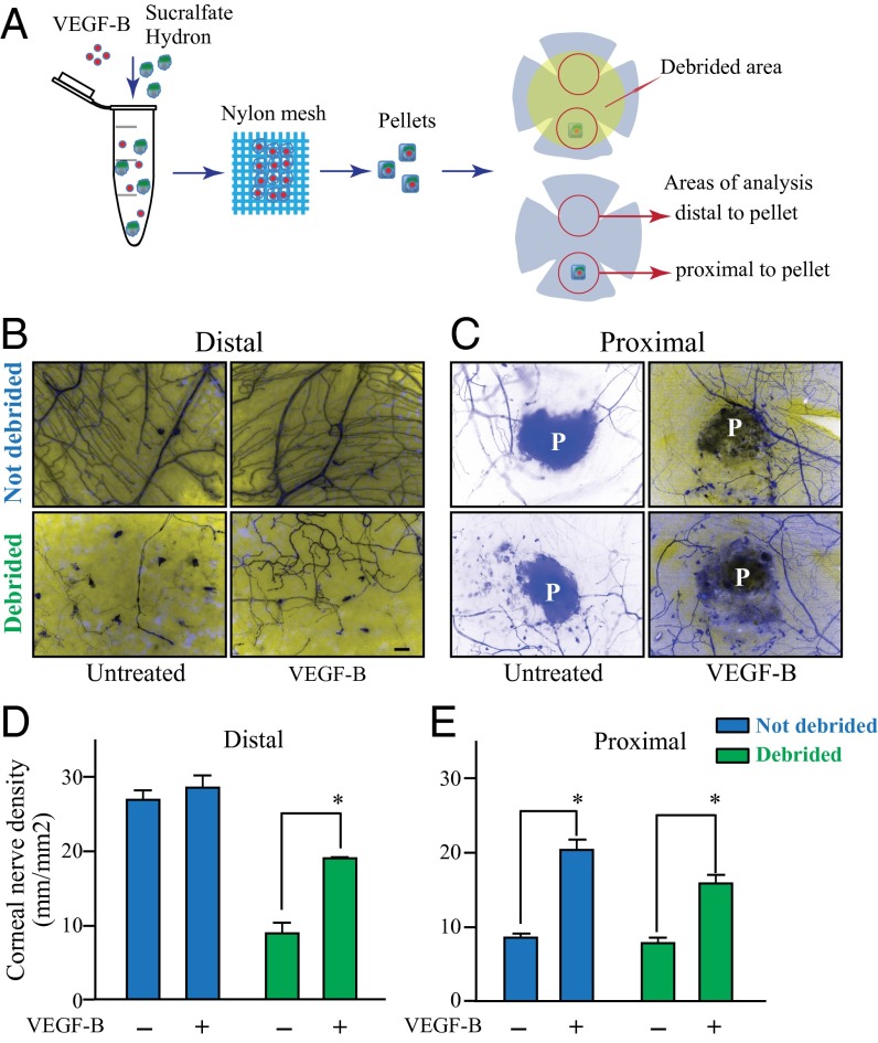
Slow release of VEGF-B into the cornea selectively enhances regeneration of injured nerves. A hydron pellet containing VEGF-B or vehicle was inserted into a corneal stroma micropocket in thy1-YFP mice. The pellet ensures the slow release of VEGF-B into the cornea. (A) One day after the procedure a subset of mice received epithelial debridement, and 3 d later corneas were collected and flat mounted for immunofluorescence analysis. Nerve growth was traced using Neurolucida software. (B, Upper and D, blue bars) In uninjured corneas, VEGF-B treatment did not induce any alteration in corneal nerve density or patterning. (C, Upper and E, blue bars) We observed a significant increase in nerve regeneration at the site of pellet implantation, which corresponded to a site of focal injury, in animals receiving VEGF-B treatment compared with vehicle controls. (B and C, Lower and D and E, green bars) When mice receiving VEGF-B–releasing pellets were also subjected to superficial corneal debridement, a more extensive injury, we observed a more profound effect, with significantly increased innervation throughout sites of injury compared with control animals. Data are expressed as mean ± SEM of two independent experiments (n = 8). *P ≤ 0.05. (Scale bar, 100 μm.)

