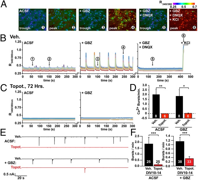
Fig. 2.
TOP1 inhibition suppresses spontaneous calcium bursts and electrical activity in cortical neuron cultures. Fura2-calcium imaging of spontaneous network activity in neurons at DIV 14–15 after the addition of 5 μM GBZ, after the addition of GBZ plus 5 μM DNQX, and after all neurons were depolarized with 55 mM KCl. (A) Representative images from vehicle-treated neurons taken at time points 1–6 indicated in B. (B) Activity in neurons treated with vehicle (Veh.). (C) Activity in neurons treated with 300 nM topotecan (Topot.) at 72 h. (D) Ca2+ burst frequency. Data are shown as means ± SEM from three independent cultures; n = 6–8 coverslips per condition; n = 466 neurons for vehicle treatment, and n = 280 neurons for topotecan treatment. *P < 0.05, **P < 0.005 (unpaired Student t test). (E and F) Spontaneous burst activity monitored by whole-cell electrophysiology in cortical neuron cultures treated with vehicle (black trace) or 300 nM topotecan (red trace). Treatment started on DIV 7 and ended on the day of recording. (E) Representative voltage-clamp recordings in ACSF and after the addition of 5 μM GBZ. (F) Burst frequency integrated from DIV 10–14. ***P < 0.0005 (significantly different from vehicle by Mann–Whitney test).

