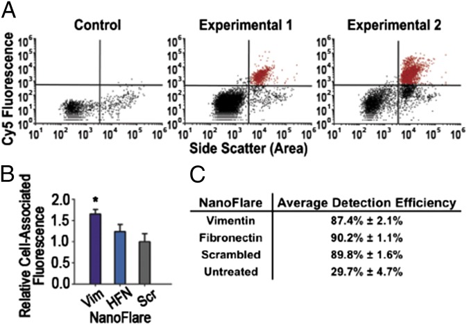
Fig. 4.
NanoFlares detect circulating breast cancer cells from whole blood in a murine model of metastatic breast cancer. (A) Representative scatter plots (n = 1 per scatter plot) show mCherry MDA-MB-231 cells recovered from whole blood by Vimentin NanoFlares from two example tumor-bearing mice (Experimental 1 and 2) (n = 12 per group) or non–tumor-bearing (Control) mice (n = 12 per group). (B) Relative cell associated fluorescence of isolated blood samples treated with Vimentin (Vim), Fibronectin (HFN), or Scrambled (Scr) Control NanoFlares added 6 h before flow-cytometric analysis. (C) Efficiency of mCherry-MDA-MB-231 cell detection by NanoFlares; values were calculated by correlating mCherry fluorescence to NanoFlare fluorescence. Untreated population detection indicates channel bleed-through of mCherry to NanoFlare channel. Samples were collected after 6.5-wk tumor inoculation. Cy5 represents NanoFlare signal. *P < 0.05.

